Often, while stroking the dog, the owner discovers a lump on its body and immediately begins to panic, suspecting that the pet has a dangerous disease. Of course, fears may not be groundless.
It is likely that the lump under the skin has a cancerous etiology, but you should not sound the alarm without showing the animal to the veterinarian, because neoplasms may be harmless and may not even require treatment.
General symptoms Usually, subcutaneous bumps do not bother dogs in any way, even if they are in the active growth phase. Depending on the cause of origin, spherical seals can have different densities and diameters - from several millimeters to several centimeters. Skin irritation and itching usually occurs with insect bites.
In other cases, the lump can only be detected tactilely. In short-haired breeds, “defects” are noticeable visually, but in long-haired breeds they are diagnosed only by palpation. If the appearance of the lump is suspicious, and the ball under the skin does not resolve within 7-10 days, then the dog must be shown to a veterinarian.
The following symptoms may alert the animal owner:
- The dog reacts painfully to palpation of the lump.
- The lump “grows” before our eyes, increasing in size several times in a short time.
- The subcutaneous tissue is opened, and pus or other discharge begins to ooze from the wound.
- The color of the skin around the lump changes.
- The pet becomes apathetic, eats poorly or refuses to eat at all, and sleeps anxiously.
Causes of subcutaneous formations
Lumps under a dog's skin are not always associated with cancer.
Most of them occur for the following reasons:
- Insect bites
. Swelling on the nose often remains after an unsuccessful encounter with bees. Their bites are always accompanied by redness and swelling. - Mechanical injury
. After an unsuccessful jump for the ball and a not at all soft landing right into the wall, a bump may appear on the face of the unlucky pet. - Infection
. With a weakened immune system, balls filled with pus often appear. This occurs due to infection by bacteria that cause inflammation. A virus can also cause compaction. - Inflammation of the paraanal glands
. If a lump is found under a dog’s tail, then its appearance is affected by the inflammatory process. The affected area will be itchy and painful. - Injection
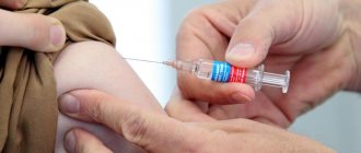
. Swelling on the thigh or paw that occurs after vaccination is a natural reaction to the injected drug. After some time it goes away on its own. - Tick infestation
. The attached parasite can be found on the stomach, side, chest, ear, chin and even under the eye. A mysterious growth that appeared shortly after a walk requires mandatory diagnosis. Some ticks carry deadly diseases.
Disorder of the functioning of the sebaceous glands and hair follicles. When the ducts are blocked, the secreted secretion accumulates inside the gland or follicle, leading to their growth.
The prolonged presence of swelling, accompanied by its increase and the appearance of pain, requires mandatory intervention by a veterinarian. Also, other alarming symptoms that are not directly related to education may be a reason for contacting.
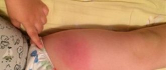
When is a bump on a dog’s withers a warning sign?
Local swelling in the injection area or a small bump after vaccination in a dog is a completely common and harmless phenomenon. Such lumps, as a rule, do not require qualified veterinary care and resolve on their own within a few weeks. In some clinical cases, when bumps on an animal’s withers are complicated by the addition of infectious microflora, the pet should be immediately shown to a doctor and adequate measures should be taken to eliminate the pathological focus of inflammation.
The dog owner should be alert to such alarming symptoms as:
- increase in size of the bump; v
- sharp redness and infiltration of the skin at the injection site;
- soreness of the lump or discomfort caused by its appearance;
- increase in local temperature after injection;
- purulent discharge from the seal;
- an increase in the dog’s general body temperature, a fever that lasts more than 3 days, a sharp deterioration in the animal’s condition, refusal to feed, apathy.
All these manifestations are regarded as pathological post-injection symptoms, and therefore require observation by a veterinarian and adequate therapy to eliminate them.
What symptoms may appear?
Not all balls appear under the dog's skin. Some of them are formed in soft tissues and can be diagnosed only by palpation of the affected area. Because of this, it is important to be aware of your symptoms.
Alarming symptoms include:
- pain when palpating the lump;
- change in color of adjacent tissues;
- stable tumor increase;
- worsening sleep, loss of activity and appetite;
- opening of the emerging papule with subsequent release of pus or blood;
- presence of temperature.
In the absence of the listed symptoms, but the formation persists for more than 7 days, it is recommended to undergo diagnostics at a veterinary clinic. If your pet has at least one of the listed symptoms, make an appointment immediately after detecting it.
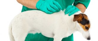
How is the diagnosis carried out?
If you notice a swelling on your dog's neck, you need to take it to the veterinarian for a thorough examination. For diagnosis, aspiration of the lump with a special thin needle is used:
- A needle is inserted into the tumor and sucks a sample of cells or fluid from the lump.
- The resulting materials are then examined under a microscope. In 90% of cases this is enough for an accurate diagnosis.
- But if studying the cells does not help the doctor make a diagnosis, then he can remove part of the lump or it completely for a detailed study of the tissue (biopsy) in the laboratory.
Did you know? According to statistics, 1 in 4 dogs over the age of 7 years develop one or another tumor.
Types of subcutaneous bumps in dogs
All subcutaneous formations are divided into 2 large groups: non-tumor and tumor. The former are often a consequence of another disease and go away after the root cause is eliminated. The latter are newly formed tissues that change healthy cells.
Benign
Benign neoplasms are characterized by the absence of pain and metastases. Despite this, over time they can develop into malignant ones, so the lack of timely diagnosis can lead to dire consequences.
Benign tumors grow very slowly or do not grow at all. This is explained by a special capsule that inhibits their increase. In most cases, they appear in a single copy and can affect any part of the body, including the animal’s finger.
Benign growths include:
- Keratoacanthomas
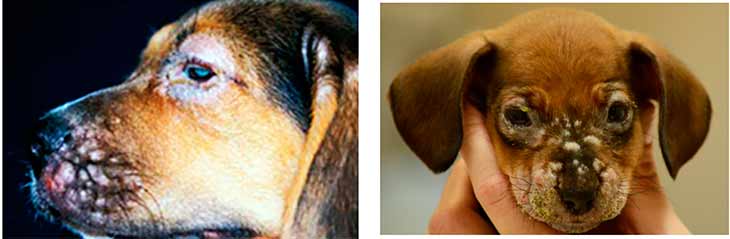
. They occur at the site of hair follicles on any part of the body covered with hair. The risk group includes shepherds. - Histiocytomas
. It often develops into an ulcer, affecting the head and ears of the animal. - Papillomas and warts
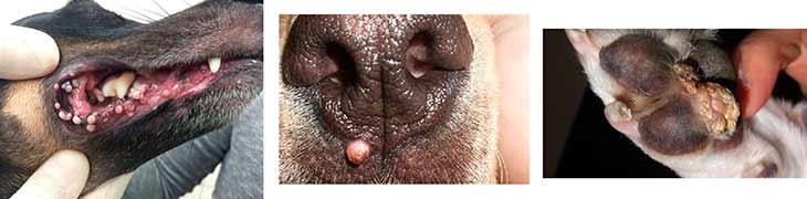
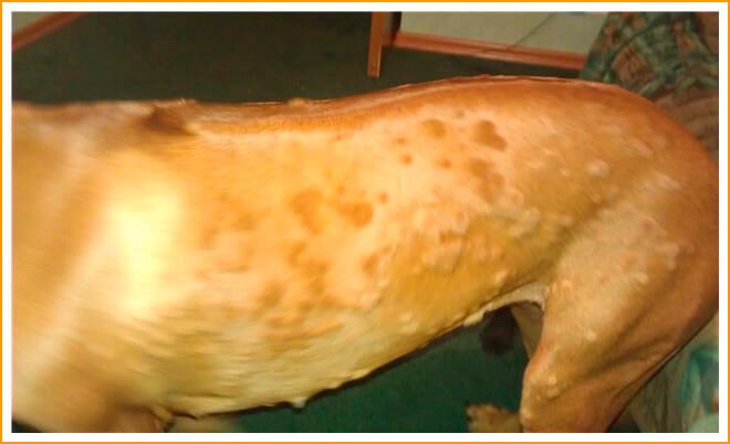
. Typical for pets with short hair. Most often, these tiny bumps appear under the skin of the dog on the back, genitals, paws and mucous membranes. They are usually caused by viral infections. - Fibroids
. Almost no different from warts and reach no more than 5 cm in diameter. Usually found on the paws. - Lipomas
. Soft, painless growths that move under the skin. An alternative name is wen. Only lipomas on the paws and carotid artery are dangerous - they can cause lameness or cause suffocation. - Hemangiomas
. They grow from the epithelial tissue of blood vessels located on the chest. Those at risk include shepherd dogs, spaniels and retrievers. - Histiocytoses
. They are formed from connective tissue in the form of subcutaneous nodules and plaques. Collies and shepherds are most often affected. - Cysts
. Loose and soft red growths are found throughout the body, including where the jaws close. Cyst-shaped balls rarely cause discomfort, so the presence of pain can be a sign of degeneration into a malignant form.
The likelihood of benign tumors developing into malignant ones is always individual. In most cases, the removal of such swellings is explained by aesthetics. Mandatory surgical intervention is recommended only for sudden changes in color, bleeding and active enlargement.
Malignant
Malignant or cancerous tumors are the result of cell mutation. Due to the lack of a restraining capsule, they quickly grow and move throughout the body, affecting it with metastases.
Cancerous tumors include:
- Lymphosarcoma
. They are formed from lymphatic tissues on the joints, under the jaws, knees and in the groin area. Occur more often than others. - Mastocytomas
. They arise from degenerated mast cells responsible for immunity. The main area of formation is the armpits, withers, groin and line along the spinal column. - Squamous cell carcinoma
. It appears as scaly plaques or red papules that transform over time into nodular formations. Such lumps, which appear under the skin of the dog on the neck or in the mouth, quickly ulcerate and penetrate into adjacent tissues. - Hemangiosarcomas
. There are cutaneous and subcutaneous types. The tumor under the skin is always black, but on the skin it can take on a bright pink tint. The subcutaneous form gives metastases in 60% of cases. The risk group includes pets with short white fur. - Osteogenic sarcomas
. One of the most dangerous neoplasms that affects bone tissue. Appear on the ribs, skull, pelvis or breast bones. The risk group includes representatives of large breeds. - Melanomas
. Round or oval spots of a dark shade that do not have a clear outline. These bumps are localized under the skin of the dog on the paws, back, eyes and mucous membranes in the mouth.
The danger of such formations lies in the absence of external symptoms at the initial stage. As the tumor metastasizes, it begins to bleed or fester. Hair falls out on the affected areas and the skin changes color. The risk group includes dogs over 8 years old.
Not tumors
In addition to bites accompanied by itching or pain, there are several other types of non-tumor formations.
These include:
- Hematomas
. Seals of various sizes that appear due to damage to blood vessels, in other words, bruises. - Pyoderma
. A purulent skin lesion often found in puppies. As a result of the disease, the body is affected by a rash or purulent papules. - Mastitis
. Inflammation of milk bags, accompanied by the formation of lumps in the nipple area. - Hernias
. Appear due to prolapse of abdominal organs. Their size varies from a pea to a chicken egg. - Lymphadenitis
. A disease of the lymph nodes that causes severe pain even with slight swelling. - Inflammation of the paraanal glands
. It clogs the excretory tract and leads to the formation of small balls resembling a hernia. - Abscess
. Purulent tissue inflammation that occurs after unsuccessful injections, injuries or infections.
The listed seals do not disrupt the functionality of healthy cells and do not change their structure. Most of these diseases do not exclude treatment, but have much less consequences than cancerous tumors.
In what cases should you consult a doctor?
Dandruff on a dog's back: causes and shampoos to get rid of
If you find a lump under your dog's skin, it needs to be palpated and examined. You should contact a veterinarian in the following cases:
- when the dog reacts painfully to palpating the lump;
- the seal changes color and increases in size;
- pus and bleeding appear;
- the animal’s temperature rises, weakness, the dog refuses to eat.
Note! To prevent your pet from licking or scratching the swollen area, you can cover the bump with a band-aid.
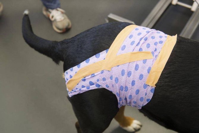
Before the doctor, you need to cover the bump with something
Most health problems can be solved by promptly contacting a veterinarian. The main thing in this case is the timely detection of a lump on the dog’s back, so you should be attentive to your pet and at least every other day, fully examine and feel your pet.
Veterinary diagnostics
A visit to the veterinary clinic is necessary if there are accompanying symptoms, a frightening appearance of the lump, or discomfort when palpating it. Before visiting the doctor, do not allow your dog to lick or scratch the affected area. This may worsen his condition.
To make a diagnosis, urine and blood tests are taken from the four-legged patient. He also undergoes X-ray, ultrasound, CT and biopsy. These studies help determine the exact size, location, depth and possible malignancy of the tumor. In the early stages, endoscopy is recommended.
What the owner should do when a lump is detected
Having discovered a lump, the owner must thoroughly wash his hands with a hygiene product, palpate from the side and above to understand how dense the texture of the formation is and whether there is something inside. This must be done carefully, as it may hurt the dog. If the lump looks suspicious, you need to take your pet to a veterinary clinic.
Important! You should not allow your dog to bite or lick the sore area. Otherwise, a granuloma will appear - a formation of connective tissue in the form of a hard knot.