What is pneumococcal infection?
Pneumococcal infection is an infection of the human body with pneumococcus, the bacterium Streptococcus pneumoniae.
In total, there are more than 90 serotypes of these bacteria. Moreover, 90% of cases of severe pneumococcal infection in children and 60% of cases of infection in adults are caused by 8 serotypes included in the pneumococcal vaccine. Bacteria do not always cause disease. A person can be a carrier of S.pneumonia and not know it. Children especially often carry pneumococcus.
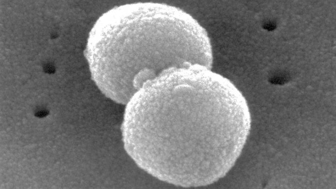
Bacteria Streptococcus pneumoniae. Photo: PHIL CDC
In kindergartens, the proportion of pneumococcal infection carriers can reach 60%, in primary school – 35%. In adults, the carriage rate is 5-7%, but when living with children it increases to 30%. Carriage implies the presence of a pathogen in the microflora of the upper respiratory tract. It is important to remember that even asymptomatic carriers can spread the infection and infect other people.
Carriage of pneumococcus can become a disease under certain conditions. This may be decreased immunity, low body resistance, hypothermia, overwork and stress. Often, pneumococcal infection becomes a complication of another disease: for example, ARVI or a cold. How the disease develops depends on the factors that provoked it. If local immunity is reduced, the infection will cause pneumonia. If bacteria enter the bloodstream, bacteremia may develop.
According to the WHO, up to 15 deaths in children under 5 years of age are associated with pneumococcus.
Effectiveness of vaccination
According to WHO, world experience has shown that mass vaccination reduces the incidence of pneumococcal meningitis and severe pneumonia in children by more than 80%, and the incidence of all pneumonia and otitis media by more than a third. The carriage of pneumococci in children is decreasing, and accordingly, unvaccinated children and adults get sick less. The World Health Organization projects that global use of pneumococcal vaccination will prevent 5.4 to 7.7 million child deaths by 2030.
Vaccination is the only highly effective way to significantly influence the morbidity and mortality of pneumococcal infections and reduce the level of antibiotic resistance in S. pneumoniae. With evidence of the safety and effectiveness of pneumococcal conjugate vaccines, WHO and UNICEF believe it is necessary to include these vaccines for children in all national immunization programs. At the same time, it should be noted that the maximum protective effect is achieved with routine vaccination of all children under 2 years of age, and not just patients in risk groups.
Transmission routes
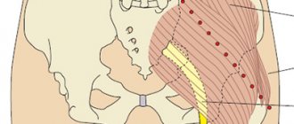
You can become infected with pneumococcus from its carrier, even if he has no symptoms of the disease. The disease is transmitted mainly by airborne droplets: during conversation, coughing or sneezing. A person easily becomes infected with pneumococcus, especially through close contact with someone who is already sick (Fig. 1).
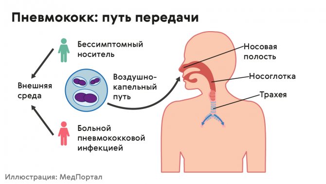
Figure 1. Routes of transmission of pneumococcus. Source: MedPortal
Who is at risk?
Anyone can become infected with pneumococcal disease. For some people, both the risk of contracting pneumococcus and the risk of severe illness due to it are higher. At risk for pneumococcus:
- Young children. The infection is especially dangerous for children under 2 years of age and often causes pneumonia, severe meningitis, and acute otitis media.
- Older people experience pneumococcal infection more severely, even if they do not have chronic diseases.
- People with bronchitis, chronic obstructive pulmonary disease, and other respiratory diseases are more likely to develop pneumonia if they become infected with pneumococcus.
- People with reduced immunity. The infection is dangerous for HIV patients, hospital patients who are undergoing long-term treatment, for people with diabetes, liver disease, cardiovascular disease, other chronic diseases, as well as for those undergoing chemotherapy for the treatment of cancer.
- Smokers. Smoking reduces local immunity, which increases the risk of infection and makes the course of the disease more severe.
Therapy for pneumococcal pneumonia
If pneumonia is mild, it is permissible to use bactericidal antibiotics orally. Phenoxymethylpenicillin, ampicillin (amoxicillin), first generation cephalosporins. If there is individual intolerance to the above drugs, erythromycin is prescribed, in some cases biseptol or its analogue groseptol. Therapy with penicillin is also acceptable. If moderate severity of pneumonia or a severe course of the disease is diagnosed, the prescription of penicillin is paramount. The drug is administered intramuscularly, 1-2 million units every 4 hours. In cases where pneumonia is complicated by pleural empyema, lung abscess, or infective endocarditis, the drugs do not penetrate the tissues very well. Then the dose of penicillin should be doubled. There are now many recognized strains of pneumococcus that are resistant to penicillin. If we are dealing with such a case, we need to prescribe cephalosporins, imipenem, vancomycin.
Diseases that can cause pneumococci
Pneumococcal infection can cause various diseases (Fig. 2), both relatively mild (otitis, community-acquired pneumonia, bronchitis) and severe, life-threatening (meningitis, bacteremia, severe pneumonia). Until 2021, pneumococcal infection caused up to 70% of the total number of pneumonias. It also causes otitis media (25% of all cases of otitis), purulent meningitis (up to 15% of cases), endocarditis and pericarditis, arthritis, sepsis and other serious illnesses.
Figure 2. Diseases caused by pneumococcus. Source: MedPortal
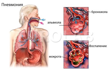
Bacterial pneumonia
Bacterial pneumonia is an inflammation of the lungs that may be accompanied by respiratory failure, fever, and severe cough. Until 2021, pneumococcal infection was the most common cause of pneumonia: it accounted for 38% of the total number of outpatient cases. Inflammation of the lungs is dangerous due to severe complications: the formation of pus in the lungs and the accumulation of fluid in the pleural cavity, the development of heart disease, secondary meningitis, and bacteremia.
X-ray of the lungs showing inflammation caused by the bacteria Streptococcus pneumoniae. Photo: Jornal brasileiro de pneumologia : publicaça̋o oficial da Sociedade Brasileira de Pneumologia e Tisilogia / Open-i (CC BY-NC 4.0)
Important! Bacterial pneumonia, caused by pneumococcus or other bacteria, is different from viral pneumonia. The viral form can develop as a complication of influenza or parainfluenza, adenoviral or new coronavirus infection. Antibiotics are not used to treat viral pneumonia; they are ineffective and can be dangerous. The exception is cases when viral pneumonia is complicated by a bacterial infection. Typically, pneumonia caused by viruses is milder and less likely to require hospitalization. For any type of pneumonia, both viral and bacterial, you must immediately consult a doctor and follow all his prescriptions and recommendations related to treatment.
Pneumococcal acute otitis media
In 30-40% of cases, acute otitis media in children is caused by pneumococcal infection. In this disease, infected fluid accumulates in the middle ear, and its mucous membrane becomes inflamed. Acute otitis media can cause hearing loss, perforation of the eardrum, and vestibular imbalance. It is also dangerous due to the spread of inflammation.
Pneumococcal meningitis
A dangerous disease in which the membranes of the brain become inflamed. Secondary meningitis occurs due to the spread of infection from other foci. Even with timely treatment of meningitis, there is a high risk of death and dangerous complications, including: hearing loss, paresis, paralysis, and mental development disorders.
Bacteremia
With bacteremia, bacteria spread through the blood, affecting organs and tissues. It can be primary or develop against the background of any other diseases associated with pneumococcal infection. Bacteremia can lead to death, especially in children (case fatality rate is 15-20%, the risk is extremely high among children with weakened immune systems and meningitis) and in the elderly (case fatality rate in this group is 30-40%).
Paranasal sinusitis
When infected with pneumococcus, the mucous membrane of the paranasal sinuses may become inflamed. Inflammation can become chronic, and abscesses, thrombophlebitis, and thrombosis can develop against its background.
Pneumococcal sepsis
A dangerous complication in which blood poisoning develops and there is a risk of severe organ damage. The risk of developing sepsis is especially high for children under 5 years of age. The condition may be manifested by a decrease in temperature, weakness and lethargy, vomiting, and inability to eat. According to WHO, the mortality rate among children with the development of pneumococcal sepsis can reach 20%.
Causes and pathogenesis of the disease
Pneumococcus most often causes inflammation of most of the lung lobe, and sometimes the entire lobe. But often pneumococcus becomes the root cause of focal pneumonia.
The medical literature describes four pathological phases of the course of lobar pneumococcal pneumonia.
1. Flushing phase, microbial swelling, redness. It is characterized by significant filling of blood vessels and strong exudation of serous fluid. Pneumococci are detected in the exudate. This phase lasts 12-72 hours.
2. Red hepatization phase. It is characterized by complete filling of the alveoli with exudate in that part of the lung that is affected by the disease. Moreover, plasma proteins (fibrinogen) are determined in the exudate, and, as a consequence of diapedesis, the number of red blood cells is increased. The area of the lung in which there is inflammation is airless, dense, acquires a reddish color and looks like the liver. The duration of this period is 1-3 days.
3. Gray hepatization phase. In this phase, significantly more leukocytes are detected in the exudate from the alveoli (mostly neutrophils), but red blood cells are significantly reduced in number. The lung, as before, is dense, grayish-yellow on the section, the granularity of the lung is very noticeable. As a result of microscopic tests, an increase in neutrophilic leukocytes and phagocytosed pneumococci is determined. This phase lasts 2-6 days.
4. Resolution phase. At this time, exudate is steadily dissolving in the alveoli. The reason for this is the influence of macrophages and leukocytes. Fibrin dissolves slowly, and the lung tissue ceases to be granular. Over time, the lung tissue regains its airiness. How long this period will last will depend on how widespread the inflammatory process is, the reactivity of the body, as well as the methods of therapy and how intensive the treatment is.
But you need to keep in mind that the disease does not always go through all these stages sequentially. Most often, in the affected lobe of the lung there are signs of several stages in combination with each other, or signs of one of the phases predominate. It is important to remember that in the case of pneumonia, pathological changes occur not only in the alveoli and interstitial tissue. The pleura, regional lymph nodes, and lymphatic vessels are affected. When diagnosed with “focal pneumonia,” the inflammation process covers a segment or lobule. At the same time, zones of compacted affected tissue alternate with areas of vicarious emphysema. Mostly serous exudate is found. It can often be purulent and contain small amounts of fibrin.
Symptoms
The course of pneumococcal infection may be asymptomatic or mild. The disease becomes dangerous if it causes pneumonia, meningitis, bacteremia or other complications. Regardless of what they are, at the beginning of the disease, severe fever with chills, shortness of breath and cough, and chest pain may appear. In older people, symptoms may be less severe, and therefore it is especially important for them to monitor their well-being and consult a doctor promptly.
The exact manifestations of pneumococcal infection depend on what kind of disease develops against its background:
- Pneumonia: temperature rises to 38-39°C, weakness, chills appear, possible muscle pain, chest pain, shortness of breath. Accompanied by a wet cough with sputum.
- Meningitis: begins with a sharp rise in temperature to 40°C and a constant headache (the sensation may be bursting). Later, vomiting and high sensitivity to light, sounds, smells and other irritants may appear. The mental state changes: a person may seem stunned, inhibited, fall into a stupor, and then into a coma. In young children with bacterial meningitis, severe irritability occurs. Convulsions may occur.
- Acute otitis media: with the disease, body temperature rises, pain appears in the ear area, sensitivity to sound stimuli becomes more acute.
- Sepsis: when the blood is infected, the body temperature rises, headaches, and weakness appear. Symptoms may depend on which organs are affected (heart, lungs, intestines, kidneys, brain, etc.). In young children, sepsis can be manifested by a decrease in body temperature, severe weakness, refusal to eat and vomiting.
Manifestations of the disease
In most cases, pneumococcal pneumonia is characterized by an acute onset and appears unexpectedly. Following a stunning single chill, a very noticeable increase in body temperature reaches 38-40°C. On the affected side, the patient feels pain while breathing. The cough is initially dry, very painful, and after a short period of time, the separation of purulent mucous sputum begins, in which blood is observed. Very often, patients notice quite a lot of such impurities - this is the so-called “rusty sputum”. Signs of intoxication in patients are very indicative, such as severe weakness, muscle and headache, almost complete lack of appetite, active tachycardia, the patient is suffocating.
Diagnostics
The main method of diagnosing pneumococcal infection is laboratory testing. Blood or cerebrospinal fluid is often taken for analysis. It is possible to study sputum, purulent discharge, fluids obtained by puncture for otitis media, sinusitis, discharge from wounds, and other biological materials. Pneumococcus is detected by microscopy, bacteriological culture, test for specific antibodies, and the presence of genetic fragments of pneumococcus. It is possible to conduct tests to determine sensitivity to antibiotics.
The doctor will conduct an examination and questioning, and evaluate the symptoms. He may order additional examinations:
- If bacterial meningitis is suspected. If a child under 5 years of age has a body temperature that rises to 38°C and there is at least one meningeal sign (impaired consciousness, increased tone and soreness of the neck muscles), a cerebrospinal fluid test is prescribed. If it is cloudy or there is leukocytosis, the diagnosis is confirmed by culture or identification of pneumococcus using PCR diagnostics and laboratory tests.
- If pneumonia is suspected. If fever, cough, or difficulty breathing occur, an x-ray or fluorography is prescribed to assess the condition of the chest organs. To confirm pneumococcal pneumonia, bacteriological culture of blood or pleural fluid is performed.
- If sepsis is suspected. Blood poisoning in adults can manifest itself as fever, deterioration in general health, shortness of breath, decreased blood pressure, and confusion. In children under 5 years of age, sepsis is manifested by a decrease in temperature, weakness, vomiting, and convulsions (the presence of at least two of these signs allows a diagnosis to be made). To damage it, bacteriological culture is performed.
Treatment
Antibacterial therapy is used to treat pneumococcal infection. It is supplemented with symptomatic treatment, which depends on the diseases caused by pneumococcus and their complications.
Important! Pneumococcus is becoming increasingly resistant to antibiotic therapy. This may make his treatment ineffective. The reason for the increase in resistance to antibiotics is their improper use, the use of medications without a doctor’s prescription, and failure to comply with the prescribed dosage or course duration. In just 7 years, from 2004 to 2011, the resistance of pneumococcal strains to antibiotics increased significantly: to penicillins from 11 to 29%, to macrolides from 7 to 26%, to co-trimoxazole from 40.8 to 50%. This worsens the treatment prognosis for those with severe pneumococcal infection.
Antibiotics are prescribed taking into account data on the resistance of pneumococcal strains common in a particular region. Beta-lactam antibiotics, macrolides, tetracyclines, and respiratory fluoroquinolones can be used for antibacterial therapy. Beta-lactam and macrolide antibiotics are preferred, but there are many common strains of pneumococcus worldwide that are resistant to them. The doctor takes this into account, as well as the peculiarities of the course of the disease, when prescribing antibacterial therapy.
For symptomatic treatment the following can be used:
- antipyretics,
- painkillers,
- non-steroidal anti-inflammatory drugs,
- antihistamines.
If bacterial pneumonia develops, the doctor may prescribe mucolytics - drugs that thin the mucus and make it easier to remove from the lungs when coughing. Bronchodilator, cardioprotective, and diuretic drugs can also be used. In parallel with antibiotics, the doctor will prescribe probiotics to maintain normal microflora of the digestive organs.
During the treatment period, it is important to maintain bed rest, drink more fluids, and eat a balanced diet. Treatment at home is only possible for mild cases of the disease. If pneumococcal infection provokes a serious illness (severe bacterial pneumonia, meningitis, bacteremia, sepsis and others), hospitalization is necessary. It is also recommended for those who are at risk: children under 5 years of age, the elderly, those with weakened immune systems and people with severe chronic diseases.
Diagnosis of pneumococcal pneumonia
Lobar pneumococcal pneumonia is characterized by certain physical manifestations, which are directly determined by the pathomorphological phase of the disease.
At the first stage of exudate accumulation, there is a dull tympanic sound above the source of inflammation, prolonged exhalation during hard breathing, and mild initial crepitus. In some cases, both types of wheezing are heard: wet and dry. During the second stage of compaction (also called hepatization), the trembling of the voice greatly increases, and bronchophony appears. During tapping, a dull sound is heard, vesicular breathing is not heard, there is no crepitus, and a noise from pleural friction is often heard. At the last stage, the trembling of the voice steadily returns to normal, bronchophony stops, redux crepitus occurs - sonorous, abundant and over a long period). In addition, the wheezing is sonorous, finely bubbled, and bronchial breathing over time turns into harsh, and then into vesicular. But it is important to take into account the fact that with pneumococcal pneumonia, these phases may not occur in the specified sequence, and in certain areas of the lungs different manifestations may be observed simultaneously. If pneumococcal pneumonia is focal, the symptoms are much less pronounced. So, in some cases, a dull percussion sound is heard above the lesion. And, as a consequence of concomitant focal bronchitis, crepitus and fine rales are heard.
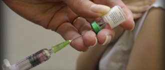
Prevention
The main way to prevent pneumococcal infection is vaccination (Fig. 3). In Russia, since 2014, vaccination against pneumococcus has been included in the National Vaccination Calendar and is carried out for children over 2 months of age. Immunization is performed with two vaccines:
- Prevenar-13. Protects against infection by 13 of the most dangerous serotypes of pneumococcus, including those resistant to antibiotic therapy. Immunization with the Prevanar-13 vaccine is carried out at 2 months, followed by revaccination at 4.5 and 15 months. The vaccine can be used for children under 5 years of age. If the first vaccination is performed later than 2 months, the doctor prescribes the vaccination regimen individually.
- Pneumo-23. Used to vaccinate people at risk: children over 2 years of age who are often and long-term ill, adults over 65 years of age, people with weakened immune systems, and people with severe chronic diseases.
The Pneumo-23 vaccine is a polysaccharide vaccine. This means that antibodies are produced as a result of a reaction to the polysaccharides that coat the pneumococcus. The bacterium itself is not included in the vaccine; it is imitated by polysaccharides that are part of its shell. The conjugate vaccine Prevenar-13 uses a carrier protein in addition to polysaccharides to form an immune response, and this speeds up the reaction and helps maintain the immunization effect longer. However, it protects against fewer serotypes of the pathogen.
Important! Children are more often vaccinated with conjugate vaccines; they form more stable immunity and protect for a longer period. Adults are vaccinated with polysaccharide vaccines; revaccination of adults against pneumococcus is carried out every 5 years.
To prevent pneumococcal infection and its complications, nonspecific measures can also be used: strengthening the immune system, taking vitamins, maintaining a healthy lifestyle, and avoiding contact with sick people.
Figure 3. Prevention of pneumococcus. Source: MedPortal
Vaccines
Vaccination has been used to combat pneumococcal infection for more than 30 years. Since 1981, the pneumococcal polysaccharide vaccine has been used. Since 2000, pneumococcal conjugate vaccines have been used in international practice for immunoprophylaxis of pneumococcal infection in young children.
To date, the following vaccines have been registered in Russia: two pneumococcal conjugate vaccines (10-valent and 13-valent - PCV10 and PCV 13) and one - polysaccharide 23-valent (PPV23). The latter is used in children over 2 years of age and adults. While conjugate vaccines are recommended for immunization of children from 2 months of age and adults aged 50 years and older. PCV13 is also registered in the US and EU for use in broader age groups (children 6 weeks to 17 years and adults 18 years and older). In the near future, the age indications for PCV13 are also expected to expand in Russia.
In 2013, the pneumococcal 13-valent conjugate vaccine Prevenar13 was awarded the Galen Prize as the best biotechnological product of the year.
More about vaccines